

Manchester, UK – 13th November 2025

Your Brain Health (YBH) has partnered with the OrthTeam Centre in Manchester, expanding access to data-driven concussion care for athletes and sports teams across the North of England – and further strengthening the UK’s national concussion care infrastructure at the forefront of digital brain health innovation.

Through the partnership, the OrthTeam Centre’s dedicated Concussion Clinic will now use ScreenIT, YBH’s digital brain health platform, to record and monitor players’ recovery through every stage of the concussion pathway. ScreenIT connects clinicians, clubs and schools through a single digital platform, creating living brain health records that make recovery measurable, trackable, and transparent – supporting safer, data-driven return-to-play decisions.

This partnership gives athletes across the North of England access to connected, evidence-based concussion care, aligning with efforts to create a consistent approach to brain health nationwide. Combining clinical expertise with real-world data helps enhance safety, recovery, and long-term brain health outcomes.

A North–South Alliance for Concussion Care

The collaboration builds on YBH’s existing partnership with Professor Mike Loosemore and the Institute of Sport, Exercise and Health (ISEH) in London – creating a north–south alliance that unites world-class clinical expertise and cutting-edge digital tools to strengthen the UK’s concussion care infrastructure.

At the heart of this alliance is a shared vision: to build a national network of “super-centres” for brain health in sport, where players can access the most advanced assessment, treatment, and monitoring – supported by a single, connected digital system.

The OrthTeam Centre’s Concussion Clinic, part of the wider Sport and Exercise Medicine Clinics network, is led by some of the UK’s most respected Sport and Exercise Medicine consultants, including Dr John Rogers, Dr Rebecca Robinson, Dr David White, Dr Jim Kerss, and Dr Bevin McCartan. It represents one of the largest multidisciplinary groups in UK sport and exercise medicine, dedicated to improving safety, recovery, and long-term brain health outcomes for athletes of all ages and abilities.

Together, YBH and the OrthTeam Centre are setting new standards for concussion management – integrating evidence-based clinical practice, connected digital infrastructure, and national collaboration to improve player welfare and long-term brain health outcomes.

Rachael Dawe, SEM Strategy Consultant at OrthTeam Centre, said: “I’m delighted by this partnership, which strengthens how concussion care is connected and delivered. By combining clinical expertise with digital innovation, we’re creating a more consistent, informed approach that supports both clinicians, patients and athletes throughout recovery.”

Dr Rebecca Robinson, Consultant in Sport and Exercise Medicine at OrthTeam Centre, said: “Your Brain Health is an asset for concussion recovery, which adds precision to the clinical approach and a digital interface at the forefront of concussion technology. Working within a multidisciplinary system with YBH and using Screen IT will enable us as clinicians to bring better recovery services to all our patients – adults and young people – experiencing concussion, and enhance how we collaborate with colleagues nationally to drive research in this important area.”

Professor Mike Loosemore MBE, Consultant in Sport and Exercise Medicine, Institute of Sport & Exercise Health (ISEH), said: “ScreenIT has transformed how we manage athletes with concussion. Its enabled seamless digital communication between clubs and the concussion clinic at ISEH, ensuring that every clinician involved has real-time access to accurate, connected data. This has improved the quality of care, enhanced collaboration, and allowed us to monitor and track recovery safely and effectively.”

David Bartlett, Chief Operating Officer at Your Brain Health, said: “Through ScreenIT, we’re building a world-leading concussion care infrastructure across the UK. This partnership with OrthTeam brings world-class clinical expertise to the North West and ensures that every player can access the best possible care. And this isn’t just for elite athletes – clubs and schools can run free baseline brain screens with ScreenIT to build a clearer picture of brain health and support safer, stronger recoveries and better-informed return-to-play decisions.”

About Your Brain Health (YBH):

Your Brain Health is a network of clinicians, researchers, and technologists dedicated to improving brain and cognitive care through advanced education, clinical support, and baseline screening. By combining state-of-the-art technology with neuroscience and clinical expertise, they’ve developed ScreenIT, their flagship platform that powers a connected digital ecosystem for proactive and precision care. This enables organisations to measure, understand, and improve brain function at scale.

About ScreenIT:

ScreenIT is an evidence-based, easy-to-use platform that supports clinicians, researchers, and communities in assessing and monitoring brain health across any setting, from the clinic to the lab to the pitch. It unifies more than 70 validated assessments spanning physical, cognitive, and psychological domains, transforming complex testing into clear, actionable insights for neurology, sports medicine, rehabilitation, and mental health, enabling precise assessment, real-time tracking, and earlier intervention.

The software has been adopted by football clubs across all levels of the football pyramid including the Premier League and WSL, Premiership Rugby sides, Franchise cricket teams, F1 teams as well as a number of Olympic Sports.

Learn more about ScreenIT

About OrthTeam Centre:

The OrthTeam Centre offers world-class expertise in musculoskeletal (MSK) care, encompassing sports and exercise medicine, sports cardiology, orthopaedics, spinal neurosurgery, peripheral nerve disorders, neurology, rheumatology, pain management, and endocrinology. Equipped with advanced imaging facilities, it provides rapid and precise diagnostics under one roof. Led by highly experienced consultants, the Centre provides expert treatment across the MSK spectrum from athletes to those with degenerative and arthritic conditions. Opened in 2019 as a joint venture with Spire Manchester Hospital, the adjacent location enables seamless transition between diagnostics and inpatient or day-case treatment.

By Associate Professor James McLoughlin

Following my colleague David Bartlett’s recent review of the Townsend et al. (2025, BJSM) paper, which quantified real-world heading forces using instrumented mouthguards across Premier League and WSL players, I turned my attention to the next logical question: how do we reduce those loads safely and effectively?

Townsend’s work confirmed that match-like drills (crosses and long balls) produce the highest rotational accelerations, and that female players consistently experience greater rotational forces than males. These findings gave us the most objective dataset yet on what happens when players head the ball. But measurement is only half the story; the real challenge lies in translating that knowledge into modifiable protective strategies.

Why Rotational Load Matters

Rotational acceleration has long been implicated as the more injurious component of head motion. Finite-element brain models show that rotational strain, particularly in cortical sulci, better predicts diffuse axonal injury and potentially chronic traumatic encephalopathy (CTE). Townsend et al. note that cumulative exposure to these rotations may predict pathology more accurately than a history of diagnosed concussions.

For clinicians, this reinforces that sub-concussive load management should focus on quality of movement, neck control, and task design, not only symptom surveillance.

The Role of Neck Strength and Control

Complementary evidence now strengthens this message.

  • Fownes-Walpole et al. (2025) combined systematic review and Delphi consensus to outline the essential components of neck-training programs for impact mitigation. Their expert panel emphasised that effective training should target:
    • Multi-planar strength and endurance
    • Dynamic stabilisation and anticipatory control
    • Sport-specific movement patterns rather than isolated static holds
  • Garrett et al. (2023, JOSPT) meta-analysed team-sport data and found a moderate negative correlation between neck strength and head-impact magnitude. Stronger necks absorb and redirect more of the incoming force, but only when activation is well-timed and directional.
  • Kavyani et al. (2025) reported that athletes with a prior concussion history demonstrate persistent neck-strength deficits, highlighting the importance of post-injury reconditioning before return to contact drills.
  • Peek (2022) provided a clear clinical framework for measurement, recommending handheld dynamometry or fixed-rig setups that capture flexion, extension, and lateral strength in neutral head posture. Reliable measurement underpins both screening and training progression.

Together, these studies shift the conversation: neck training is not an optional extra, but a primary prevention and rehabilitation strategy for athletes exposed to repetitive head loads.

Technique and Tactical Preparation

Prevention also extends beyond musculature.

  • Peek et al. (2025) urged a “re-think” of head-injury prevention through tactics and technique. Coaching points such as body positioning, timing of jump, and angle of approach can meaningfully alter both impact location and rotational torque.
  • Ross et al. (2025, HeaderPrep) demonstrated that targeted heading-readiness programs for youth female players are both feasible and well-accepted, improving confidence and technique while limiting high-force exposures.

For practitioners, these findings support a progression model: prepare before exposure. Blending neuromuscular control, technical education, and measured load increments.

Translating Evidence Into Practice

Quantify and Monitor

Whenever possible, use objective measures such as validated digital tools like instrumented mouthguards or video coding to track exposure patterns over time. Even periodic sampling can highlight positional or drill-specific risk.

Structure Heading Drills

  • Begin with low-velocity, “thrown” headers, focusing on timing and neck control.
  • Progress to aerial crosses and long-ball scenarios only once mechanics and anticipatory activation are stable.
  • Limit overall high-force exposures, particularly across congested training weeks or in younger players.

Integrate Neck Training Year-Round

  • Combine isometric holds, dynamic perturbation exercises, and multi-directional resistance (e.g., band or partner drills).
  • Train in football-relevant postures: semi-flexed trunk, reactive stance, rather than supine positions.
  • Review progress every 4–6 weeks using consistent testing positions.

Educate and Communicate

Ensure players understand why load management matters. Encourage disclosure of dizziness, neck fatigue, or delayed headache after repetitive headers, symptoms that can reflect both musculoskeletal and vestibular strain.

Implications for Female and Youth Athletes

Townsend et al. found higher rotational loads in female players, aligning with other data showing increased concussion incidence in women’s football. Potential contributors include lower baseline neck strength, smaller head-to-ball mass ratios, and different heading mechanics.
Clinicians should therefore:

  • Establish sex-specific baselines for neck strength and control.
  • Introduce graduated “header readiness” programs for adolescent and female players before exposure to match-like drills.
  • Advocate for equitable inclusion in future research. Female cohorts remain markedly under-represented.

The Bigger Picture

Collectively, these studies provide the framework football has long needed:

  • Townsend 2025 quantifies how much and how hard players head the ball.
  • Fownes-Walpole, Garrett, and Kavyani explain how the neck contributes to mitigating load.
  • Peek and Ross show how to coach and measure it in real settings.

For clinicians, this convergence of evidence allows more precise conversations with coaches, strength staff, and governing bodies about “smart exposure” — protecting brain health without losing the skill of heading.

Take-Home Summary

Focus Area Practical Action
Load monitoring Use validated tools (iMGs, video,) to quantify session frequency and intensity.
Neck conditioning Integrate progressive, multi-directional, task-specific training 2–3× per week in preseason, then maintain weekly. Screen neck measures
Technical coaching Teach timing, posture, and ball-flight anticipation to reduce rotational acceleration.
Player education Emphasise cumulative risk and the role of fatigue; encourage early reporting of neck or vestibular symptoms.
Female/youth focus Lower exposure thresholds; prioritise skill and readiness over repetition.

Closing Thought

As Townsend et al. conclude, the aim is not to eliminate heading but to guide it. With a deeper biomechanical understanding, targeted neck-training protocols, and modern monitoring technology, clinicians can lead football toward a future where every header is both skilful and safe.

References

Townsend, D. C., Jones, C., Patel, S., Green, M., Riley, P., Brownlow, M., Gillett, M., & Belli, A. (2025). Heading to guidance: Understanding in-training heading demands for elite men’s and women’s football. British Journal of Sports Medicine. Advance online publication. https://doi.org/10.1136/bjsports-2024-109525


Fownes-Walpole, M., Heyward, O., Till, K., Mackay, L., Stodter, A., Al-Dawoud, M., Bussey, M. D., Gordon, L., Hairsine, J., Kirk, C., Madden, R., McBride, L., McDaniel, A., McKnight, P., Mill, N., Peek, K., Pratt, G., Ryan, D., Salmon, D., … Jones, B. (2025). Combining evidence and practice to optimise neck training aimed at reducing head acceleration events in sport: A systematic review and Delphi-consensus study. British Journal of Sports Medicine. Advance online publication. https://doi.org/10.1136/bjsports-2024-108847


Garrett, J. M., Mastrorocco, M., Peek, K., van den Hoek, D. J., & McGuckian, T. B. (2023). The relationship between neck strength and sports-related concussion in team sports: A systematic review with meta-analysis. Journal of Orthopaedic & Sports Physical Therapy, 53(10), 1–9. https://doi.org/10.2519/jospt.2023.xxx


Kavyani, A., Bourne, M., Williams, M., Timmins, R., Peek, K., Bennett, H., Mastrorocco, M., & Garrett, J. M. (2025). The impact of sport-related concussion history on neck strength in elite Australian rules football and rugby league athletes. Physical Therapy in Sport. Advance online publication. https://doi.org/10.1016/j.ptsp.2025.08.001


Peek, K. (2022). The measurement of neck strength: A guide for sports medicine clinicians. Physical Therapy in Sport, 55, 282–288. https://doi.org/10.1016/j.ptsp.2022.04.003


Peek, K., Georgieva, J., Wilson, B., Massey, A., & Serner, A. (2025). Re-thinking head injury prevention in football: The role of tactics and technique. Journal of Science and Medicine in Sport. Advance online publication. https://doi.org/10.1016/j.jsams.2025.07.009


Ross, A. G., Whalan, M., Duffield, R., & Peek, K. (2025). Can we prepare young female players for heading in football? The feasibility and acceptability of HeaderPrep. Journal of Science and Medicine in Sport, 28(11), 894–899. https://doi.org/10.1016/j.jsams.2025.05.014

By David Bartlett, Physiotherapist in the Concussion Clinic at Institute of Sport & Exercise Health (ISEH)

The recent British Journal of Sports Medicine paper by Townsend et al. (2025) marks a significant step forward in our understanding of heading exposure during football training. For the first time, elite male and female footballers were monitored using instrumented mouthguards (iMGs) to capture the real-world frequency and intensity of headers, moving beyond laboratory estimates and self-report data that have long limited this area of research.

Across 63 professional training sessions, the study recorded nearly 1,500 heading events. The results revealed average peak linear accelerations of 18 g and rotational accelerations of ~1,000 rad/s², with female players consistently experiencing higher rotational accelerations than males. Crucially, match-like scenarios such as crosses and long balls produced the highest forces, while throw-ins, more common in training drills, resulted in lower impacts.

A step forward for football science

This research represents tangible progress. The methodology adheres to the Consensus Head Acceleration Measurement Practices (CHAMP) framework, using a validated iMG technology (Protecht) to produce the most reliable dataset yet on heading in elite football. It provides an evidence base for training-load management and begins to inform guidance on limiting repetitive head impacts, a necessary foundation for future policy and practice.

The use of wearable technology across both men’s and women’s elite tiers should be recognised as a milestone for player welfare. For the first time, we can meaningfully quantify what a “typical” training exposure looks like, rather than relying on conjecture or extrapolation from match data.

But peak force understanding remains limited

While the study provides robust quantification of how often and how hard players head the ball, it stops short of answering the critical question; what do these forces mean for the brain?

The peak linear and rotational accelerations recorded remain well below concussive thresholds, yet our understanding of the cumulative or sub-concussive impact of repetitive exposure remains incomplete. Rotational acceleration, in particular, is thought to exert greater strain on neural tissues, but the clinical consequence of these training-related exposures remains speculative.

This is especially relevant for women’s football, where the study identified significantly higher rotational accelerations but could not determine why. Possible explanations include differences in neck strength, head-neck segment mass, or heading technique, all of which demand closer biomechanical and neuromuscular scrutiny.

The practical takeaway: neck strength and neuromuscular control matter

What this study reinforces, perhaps more than anything, is the need for targeted cervical spine conditioning as part of concussion-prevention and performance programmes.

Strong, well-coordinated neck musculature could reduce head acceleration by stabilising the head–neck complex at the moment of impact. In practical terms, this means progressive strength and proprioceptive training, ideally integrated into existing strength and conditioning or physiotherapy routines.

For female athletes, who may be more susceptible to higher rotational forces, this may carry even greater importance. Tailored neuromuscular interventions that improve timing, co-contraction, and dynamic control could be key to mitigating risk without compromising performance.

Progress made

Townsend et al. should be commended for delivering the most comprehensive quantification of training-related heading to date. Their findings are a clear advance in the ongoing effort to understand, and ultimately manage, the neurological load placed on footballers.

But quantification is not the same as comprehension. Until we better understand how these forces translate into brain strain, metabolism, and long-term neurodegenerative risk, our response must combine data-driven exposure management with proactive neck-control conditioning.

In short: progress has been made, but the science of protection is only just beginning.

View Video

Jordan Cox’s stunning catch to dismiss Steve Smith: a masterclass in gaze stability.

By David Bartlett, Physiotherapist at Welsh Fire


When Jordan Cox sprinted toward the boundary and plucked a soaring ball to dismiss Steve Smith in the Hundred this summer, the crowd saw a moment of pure athletic brilliance.
Those of us working at the intersection of brain health and performance saw something more: a live demonstration of world-class gaze stability.

The Invisible Work Behind the Catch

Tracking a cricket ball that is descending at over 100 km/h while your own body is accelerating is a neuromechanical challenge of the highest order. As Cox turned and ran back, his cervical spine moved from deep extension and left rotation to a neutral posture, all while the visual backdrop shifted abruptly from the uniform blue of the sky to the high-contrast chaos of a packed grandstand.

For the ball to remain sharply focused on his fovea, Cox’s vestibulo-ocular reflex (VOR), cervico-ocular reflex (COR) and smooth pursuit eye tracking functions all had to perform flawlessly. These oculomotor functions integrate information from semicircular canals of the inner ear, neck muscle spindles, and joints to control extra-ocular muscles, driving equal-and-opposite eye movements within roughly ten milliseconds of head motion. Any deficit in gaze stability gain, even mild, would have produced compensatory corrective saccades, causing the ball to blur or “jump” in his visual field. In that scenario, the catch simply doesn’t happen.

Implications for Performance

This is where what we know from concussion management and performance science needs to converge. We know from both clinical research and daily practice that even subtle vestibular, cervical and oculomotor impairments after head trauma degrades oculomotor functions and dynamic visual acuity. Cervico-vestibular dysfunction, common after rapid head acceleration injuries, together with physiological injury to brain and brainstem pathways, adds another layer, as proprioceptive input from the neck is essential for accurate gaze control.

Yet traditional return-to-play assessments often stop at symptom checklists or static balance tests. Cox’s catch is a compelling reminder that sport demands far more. If an athlete cannot maintain visual clarity while sprinting, rotating, and reacting to a shifting background, they are not truly match-ready.

Training and Screening the Invisible System

The good news is that gaze stability can be trained and measured. Dynamic visual-acuity tests, head-impulse assessments, oculomotor tests and progressive vestibular rehabilitation (the classic X1 and X2 drills, for example) and sports specific gaze stability exercises provide both objective metrics and effective interventions. Embedding these in pre-season screening and post-concussion protocols should now be as routine as hamstring strength testing.

A Broader Lesson

What fans celebrated as a spectacular dismissal was, at its core, a triumph of incredible neuro-ocular-vestibular-cervical integration. For performance and medical teams, it highlights a simple but critical truth: protecting and optimising the brain–eye–neck axis is not a niche clinical concern, it is a competitive necessity.

Elite catches are born not only of athletic talent but of a nervous system tuned to keep a stable gaze on moving targets, while the body moves at speed. In professional sport, that is as worthy of training and safeguarding as any other physical skill.

By Associate Professor James McLoughlin 

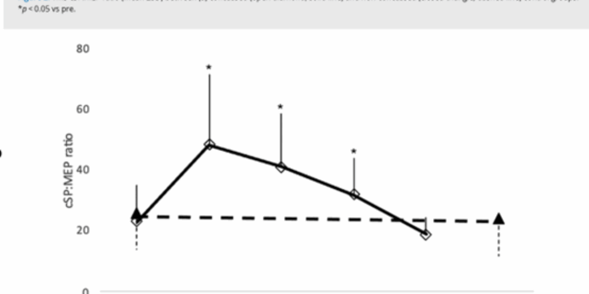
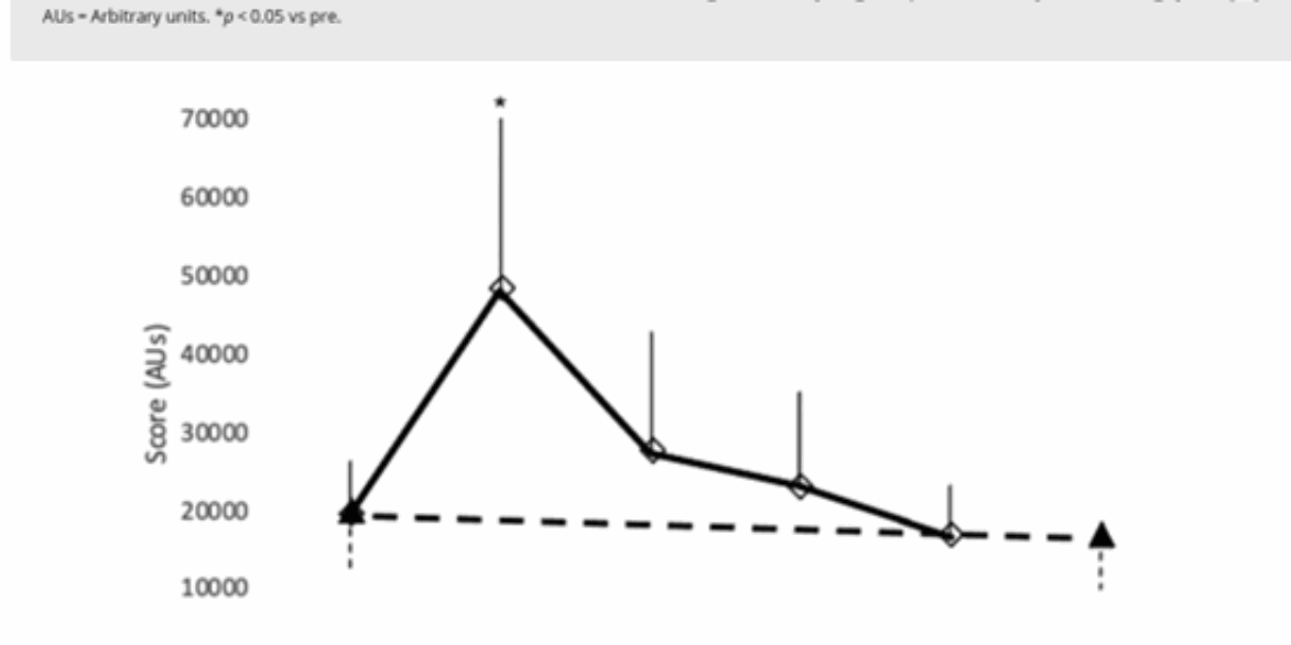
The recent Herald Sun article1, based on a new Swinburne University study using transcranial magnetic stimulation (TMS)2, highlights what many clinicians have long recognised: concussion recovery is not as simple as counting down the days.

In sport, return-to-play (RTP) rules are often based on arbitrary timelines; 12 days in elite AFL, 21 days in community levels, rather than an accurate picture of brain recovery. This study found that while athletes reported feeling symptom-free after about 12 days, measures of cortical inhibition (via TMS) were still abnormal for up to 26 days.

Every day in Australia, we hear that an athlete is sidelined due to “concussion protocols”, rather than what is really happening; the athlete is recovering from a concussion injury. Concussion is a rapid head acceleration injury with neurophysiological, musculoskeletal, and psychological consequences. Just like a hamstring or shoulder injury, recovery requires an individualised, multimodal assessment, not an arbitrary timeline. You don’t hear of players out with “hamstring protocols”, they are recovering from a hamstring injury. And running without pain symptoms certainly does not mean you have fully recovered!

This reinforces what we see clinically every week: symptom resolution does not necessarily mean full recovery.

Research Tools vs. Clinical Reality

I have followed TMS research closely for over two decades. It provides fascinating insights into brain physiology, but I remain cautious about its role in clinical decision-making. The same caution applies to other emerging research tools, like visual evoked potentials (VEPs), which are currently being pitched as clinical aids but are still largely investigational3.

These technologies give us another lens into brain recovery, but the reality is that a comprehensive clinical assessment still provides the most meaningful guidance for safe RTP decisions.

Interestingly, this very study also included a smooth pursuit oculomotor test and the recovery timelines it revealed were remarkably like those detected by TMS (see Figure 1 & 2)2. This illustrates that clinical tools already in use can provide equivalent and actionable insights.

Figure 1

Figure 2

The Power of Multimodal Clinical Screening

Within our global Your Brain Health (YBH) network, oculomotor smooth pursuit testing is already routine. But it’s just one piece of a much larger puzzle. Clinicians in our network use a multimodal screen that assesses:

  • Smooth pursuit, saccades, anti-saccades, gaze stability, and optokinetic movements with Neuroflex®.
  • Objective measures of postural sway, balance and vestibular signs & symptoms
  • Cognitive functions such as attention, memory, and processing speed
  • Mental health, sleep, and confidence
  • Coordination and broader motor control, including cervical spine contributions
  • Relevant medical history and contextual factors

This multimodal approach provides a clear, real-world picture of recovery—and directly informs decisions about returning to play, learn, and work. The idea that symptom recovery alone does not align with objective markers is not new. In fact, defining recovery purely in terms of symptom resolution has been one of the greatest flaws in concussion research to date.

Why This Matters

Every athlete deserves recovery care that is:

  • Individualised – no two concussions recover the same way
  • Comprehensive – covering brain, body, and psychological health
  • Transparent – so players, families, and clinicians can track progress together

This is one of the reasons we built ScreenIT, software that integrates all these clinical measures, streamlining care for the individual while also creating robust, longitudinal datasets to advance concussion research.

Moving Forward

The Swinburne study is important because it reinforces that relying on symptoms—or arbitrary timeframes—alone is not enough. But rather than waiting for research tools like TMS or VEPs to become clinically viable, we already have the ability to perform comprehensive, multimodal assessments that guide safer recovery today.

By combining best-practice measures with emerging technologies, we can ensure athletes return not just when they feel ready, but when their brains and bodies are truly ready to perform at their best.

Bottom line: Concussion recovery should not be dictated by the calendar. Neurophysiological tools like TMS and VEP provide useful insights, but the cornerstone remains a comprehensive, multimodal clinical assessment—supported by technology like ScreenIT and delivered by skilled clinicians who understand the complexity of brain health.

References

  1. Clarke, B. (2025, August 17). When do you recover from a concussion? Shock new findings. Herald Sun. https://www.heraldsun.com.au/health/mental-health/concussion-recovery-periods-may-be-too-short-new-brain-study-suggests/news-story/73aa4cf85aa55e875b42c3497f96651b
  2. Pearce, A. J., Middleton, K., & Clarke, A. (2025). Time-course responses following sports-related concussion: a multi-modality study. The Physician and Sportsmedicine, 00913847.2025.2541579, 1–9.
  3. Fong, D. H. C., Cohen, A., Boughton, P., Raftos, P., Herrera, J. E., Simon, N. G., & Putrino, D. (2020). Steady-state visual-evoked potentials as a biomarker for concussion: A pilot study. Frontiers in Neuroscience, 14, 171.

By Associate Professor James McLoughlin 

Over the past decade, vestibular education has strongly emphasized the role of the Head Impulse Test (HIT) and its video-based cousin (vHIT), particularly in acute settings. This focus stems from their pivotal role in the Head Impulse, Nystagmus, Test of Skew (HINTS) protocol, which—when applied accurately and in the right context—can help differentiate central causes (e.g., stroke) from peripheral vestibulopathies (e.g., vestibular neuritis). Rightly so: it’s a powerful, bedside decision tool in emergency neurology. 

However, I have noticed over the past 5 years, this stroke-centric application of HIT/vHIT taught in many vestibular courses has disproportionately shaped the broader clinical conversation—especially in rehabilitation and sports medicine. Too often, clinicians are left with the impression that a normal vHIT rules out significant dysfunction. In reality, this is where functional vestibular assessment should begin. 

The Limits of HIT/vHIT 

HIT and vHIT primarily assess high-frequency, high-acceleration components of the vestibulo-ocular reflex (VOR). They’re excellent at detecting large, acute deficits in semicircular canal output. But these tools do not capture: 

  • Low- and mid-frequency impairments 
  • Central integration deficits 
  • Dynamic visual acuity 
  • Symptom provocation during movement 

In cases like concussion, cerebellar ataxia, migraine-associated dizziness, or motion sensitivity, the vHIT may be entirely normal while patients still report disabling dizziness, fogginess, or blurred vision during head movement. 

Functional Gaze Stability = Everyday Brain Performance 

Patients don’t live in a vHIT lab. They live in dynamic environments—navigating busy streets, scanning playing fields, or walking through supermarkets. These real-world tasks require gaze stability across a variety of head speeds, directions, and cognitive loads. 

We must assess gaze stability across a range of speeds and tasks to: 

  • Identify direction and speed specific subtle deficits 
  • Track rehab progress 
  • Assessing cervical-vestibular coordination and compensatory strategies 
  • Tailor VOR retraining 
  • Guide return-to-play and return-to-learn decisions 

Even simple tools like Dynamic Visual Acuity (DVA) and the VOMS battery can reveal critical deficits missed by vHIT. 

Concussion and Cerebellar Cases 

In concussion, vestibular symptoms often reflect central processing issues, not peripheral loss. Patients may pass vHIT yet experience visual blurring, dizziness, or cognitive fatigue. 

Cerebellar disorders affect the coordination of eye-head movement and often require dynamic, functionally relevant testing to identify deficits. 

Recalibrating Our Focus 

vHIT is a starting point. To support recovery, clinicians could incorporate: 

  • Smooth pursuit and VOR cancellation 
  • DVA at varied speeds 
  • Head precision and proprioceptive control (e.g., HeadX Kross) 
  • Functional movement with gaze tasks 

Final Thought 

If symptoms persist, dig deeper than just HIT and vHIT. Gaze stability is not binary. Like all brain functions, it must be assessed across varied speeds, loads, and contexts to understand and treat it most effectively. 


Adelaide, 2 July 2025

Your Brain Health’s flagship digital platform, ScreenIT, has been awarded top honours in the Technology Platform category at the 2025 South Australian iAwards, held at KPMG Adelaide on 1 July.

Hosted as part of the 32nd annual AIIA iAwards, the event celebrated innovation excellence across the state. South Australia’s Assistant Minister for Artificial Intelligence and the Digital Economy, Michael Brown MP, addressed attendees, highlighting the transformative potential of AI for economic growth and the importance of industry collaboration in managing emerging technologies responsibly.

Developed in close partnership with MPF, ScreenIT is redefining how clinicians and elite sports organisations manage brain health. With its intuitive interface and integrated SCAT6/SCOAT6 tools, the platform is already empowering teams to make faster, smarter decisions around concussion and cognitive recovery.

“This award recognises the power of purpose-built technology to improve health outcomes and streamline clinical workflows,” said Greg Rosenbauer, CEO of Your Brain Health. “We’re thrilled to see ScreenIT acknowledged for its impact, and excited for what’s ahead.”

The win qualifies ScreenIT for the national iAwards, to be held in August.

Learn more about ScreenIT →

By David BartlettChief Operating Officer at Your Brain Health and Welsh Fire Physiotherapist.

When an athlete sustains a potential concussion, the decisions made in the moments that follow are critical. Healthcare Professionals and medical staff are often required to act quickly, using the best tools available to assess injury severity and guide next steps. The SCAT6 (Sport Concussion Assessment Tool, 6th Edition) has emerged as the leading sideline and clinical evaluation tool—but is it truly the best we have?

The Evolution of Concussion Assessment

Concussion assessment has come a long way. Decades ago, decisions were based largely on observation and subjective judgment. The introduction of structured tools like the SCAT series from the Concussion in Sport Group (CISG) transformed this landscape—bringing standardised protocols, objective criteria, and evidence-informed decision-making into both sideline and clinical environments.

The latest iteration from the 2022 consensus meeting, SCAT6, incorporates years of research and expert feedback. It’s no longer just a checklist; it’s a comprehensive, multi-domain assessment tool covering symptoms, cognitive and neurological function, balance, and more. Updates like the 10-word recall task address previous concerns about sensitivity, particularly in elite athletes.

The Strengths of SCAT6

SCAT6 is widely used as the go-to concussion assessment tool for the acute phase of injury. It encourages a holistic view of the athlete, prompting clinicians to consider symptom progression, cognitive status, and observable signs rather than relying on any one domain.

It also reinforces a culture of athlete safety. The emphasis on standardised removal-from-play protocols and serial assessments helps mitigate the risks associated with premature return-to-play decisions.

Persistent Limitations

Despite its strengths, SCAT6 is not without limitations. It remains a tool—not a diagnostic verdict—and is dependent on the clinical judgment and skill of the person administering it. Its validated window of utility is confined to the first 72 hours post-injury, limiting its role in ongoing management.

That distinction—tool, not diagnosis—is crucial, yet often misunderstood. Many research studies still use SCAT6 outcomes as de facto diagnostic criteria for inclusion or exclusion, despite the fact that SCAT6 was never intended to serve as a standalone diagnostic standard. This creates inconsistencies in the literature and can contribute to overreliance on thresholds that were meant to guide, not define, clinical decision-making.

Practical barriers also persist. The tool’s comprehensiveness can be a double-edged sword in time-pressured environments. Administering SCAT6 efficiently requires training, familiarity, and ideally, baseline data—which not all settings can support.

Historically, one of the major constraints has been the lack of a digital format. Paper-based assessments can be cumbersome, prone to inconsistencies, and difficult to integrate into broader EMR systems or performance tracking platforms.

That’s why the arrival of a digital SCAT6 is a significant step forward. Digitisation streamlines administration, enables structured data capture, supports longitudinal tracking, and reduces variability in how the tool is used across clinicians and environments. In real-world terms, this means faster workflows, cleaner data, and more reliable follow-up over time.

Where Does SCAT6 Fit Within the Broader Landscape?

SCAT6 offers a robust and integrative approach, integrating other tools—such as the Standardised Assessment of Concussion (SAC), Balance Error Scoring System (BESS), and simple symptom checklists. However, it is not designed to function in isolation. It should be seen as part of a clinician’s broader toolkit, not a substitute for comprehensive evaluation or clinical reasoning.

It’s also important to acknowledge gaps in evidence for certain populations—children under 13, para athletes, and female athletes, for example—where normative data is still evolving. Ongoing research and future iterations of the SCAT will need to address these limitations more fully, although in 2022 the CISG also released the Child SCAT6.

A Tool That Keeps Improving

So, is SCAT6 the best we have? For the acute assessment of sport-related concussion, it is certainly the most complete tool currently available. And with the shift to digital formats, some of its practical limitations are being actively addressed—making it more accessible, efficient, and clinically useful.

But like any tool, its effectiveness depends on context, competence, and careful application. SCAT6 should support—not replace—clinical judgment. As our understanding of concussion continues to evolve, so too must our tools and technologies.

For now, SCAT6 stands at the forefront of concussion assessment. And with modern platforms like ScreenIT offering digitally integrated SCAT6, we’re better positioned than ever to provide timely, informed, and athlete-centered care.

By Associate Professor James McLoughlin, Chief Academic Officer at Your Brain Health

Step 1: Confirming the Concussion

Think of this as saying, “Yep, you’ve had a knock.” It’s an important first step — but it’s only the beginning.

Imagine a car that’s been in a minor crash. The first thing you do is check for visible damage. Confirming a concussion is similar. The brain — along with the neck and brainstem — has taken a hit, and there’s been a temporary change in function.

In the immediate aftermath, the top priority is determining whether emergency medical care is needed. We look for red flags that require an immediate medical response. Following this we monitor physical, cognitive, and emotional symptoms over the next few days. This process should be overseen by a responsible adult — not your mates while out at the pub.

To help guide decision making in this acute phase, the Concussion Recognition Tool 6 (CRT6) is the go-to resource. It’s simple, safe, and designed for use by coaches, trainers, parents, and anyone involved in player care. It helps recognise red flags and core symptoms and provides helpful advice for what to do next.

Yes, emerging technologies like blood biomarkers, saliva tests, and wearable sensors are exciting — but they need to add value. That means improving decisions and guiding actions. These tools must be co-designed with those on the front lines: players, physios, coaches, and carers. Plenty of apps and other portable measures of specific brain functions are now hitting the market. However, if it doesn’t support and enhance decision-making, it’s not helping.

Step 2: Profiling Brain Health

This is where concussion care gets truly clever.

Knowing someone has had a concussion is one thing. But understanding how it’s affecting them is another. Is balance off? Vision blurry? Thinking slow? Mood unstable? Sleep disrupted? A bit of everything?

Now we’re popping the hood to see what’s really going on.

Multimodal brain profiling goes beyond diagnosis. It assesses the systems most often disrupted by concussion, including:

  • Symptoms (e.g. headache, dizziness, nausea)
  • Mental health
  • Sleep quality
  • Vestibular system function
  • Cervical spine function
  • Oculomotor function
  • Balance and coordination
  • Cognitive performance
  • Autonomic nervous system regulation

To get the most accurate picture, we also integrate:

  • Individual brain health history
  • Previous baseline brain health screening data

Without evaluating all of these domains, you risk missing key information. But by using comprehensive brain health profiling, we can track recovery, guide referrals to the right professionals, and tailor rehabilitation to the individual’s needs.

Excitingly, repeatable baseline screens now allow us to track these domains over time. This opens the door to a more preventative and personalised approach to brain health — targeting modifiable risk factors long before issues become chronic. Multimodal brain health profiling, built into platforms like ScreenIT, enables clinicians to deliver this personalised approach at scale. And with nearly half of all dementia cases considered preventable, this kind of proactive strategy is a genuine game-changer.

Why It Matters

When we know which systems are affected, we can deliver targeted support — whether it’s neck physiotherapy, balance training, vision rehab, heart rate-guided aerobic exercise, or structured rest strategies.

We’re not just managing the concussion — we’re tuning the whole system. That includes identifying pre-existing conditions (e.g. migraine, anxiety, ADHD) that might influence how we approach rehabilitation and recovery.

This leads to:

  • Faster, safer return to activity
  • Reduced risk of prolonged symptoms
  • Better outcomes across the board

So, the next time you hear about a “new tool” to diagnose concussion, ask: Does it help improve care? Does it inform recovery planning?

Diagnosis is step one. But multimodal brain health profiling continues to evolve — and it’s here to stay!

By Associate Professor James McLoughlin, Chief Academic Officer at Your Brain Health.

This week, I’ve been fielding a lot of questions about the recent Herald Sun coverage (also picked up in the Adelaide Now) on a new concussion detection tool, BrainEye. You can read the article here:

🔗 BrainEye app delivers 100% concussion detection rate 

 Naturally, I was curious to dive into the new research backing this product: 

 Clough, M., Bartholomew, J., White, O., & Fielding, J. (2025)
Investigating the utility of the BrainEye smartphone eye tracking application and platform in concussion management. Sports Medicine – Open, 11(1), 24. 

The Study at a Glance 

The research involved 384 AFL players from 10 of the 18 clubs, using BrainEye for baseline testing. However, only three clubs provided post-concussion data. That left: 

  • 11 players tested on the day of concussion 
  • 9 tested at 48 hours 
  • 5 tested at 7 days 

BrainEye tested four eye movement and pupil functions:  

  • Smooth Pursuit (SMP) 
  • Near Point Convergence (NPC) 
  • Pupillary Light Reflex (PLR) 
  • Horizontal Gaze Nystagmus (HGN) 

Only two tests—SMP and PLR—produced usable data. This didn’t come as a surprise to us, based on our own early look at the device. 

The results? The combined BrainEye score had a reported 100% sensitivity and 85% specificity for identifying clinical concussion in this small group. Individually, SMP and PLR showed moderate diagnostic value, with sensitivities around 70%. 

While that all sounds impressive, especially to a headline editor, it’s worth taking a closer look at what this actually means for real-world application. 

Optimism vs. Reality: The Big Picture 

Let’s start with what’s promising: 

  • Objective data in eye movement and pupil response is increasingly recognised as a key part of the concussion puzzle. 
  • Having rapid, accessible tools could help in environments where expert clinicians aren’t available pitchside. 
  • BrainEye’s attempt to move this tech onto a mobile device is a logical step forward in usability. 

But here’s the catch: 

  • The study only tracked 11 concussed players, all of whom were already clinically diagnosed using standard tools like SCAT6. So, the real-world added value of BrainEye in this context remains unclear. 
  • Two of the four test domains weren’t usable in this setup. 
  • It’s not yet known if the smartphone-only app (without the infrared hardware used in the study) can deliver the same data quality. 
  • Implementation was low—just 3 of 10 clubs actually used the tool post-concussion, citing logistical barriers. 

The Grassroots Question 

A key concern is how this technology is being positioned. The research is based on a small, elite-level AFL sample, but the marketing is targeting grassroots players and junior leagues. Is the tool validated in that context? Does it actually help detect concussion better than tools like CRT6 (used by non-medical professionals)? 

If the goal is broader safety, we need to ask: 

  • Does it lead to more players being correctly removed from play and referred for medical evaluation? 
  • Can it inform recovery, not just detection? 
  • Does it support targeted vestibular, visual or cervical rehab pathways? 

These are the real-world questions that matter—not just headline accuracy claims from a pilot study. 

Where We Stand at Your Brain Health 

At Your Brain Health, we’ve been using several high-resolution, oculomotor tests as part of a multimodal brain health screen for over three years. And that’s just one part of our toolkit. 

We’re not just assessing injury—we’re tracking recovery and guiding referral pathways for: 

  • Vision and vestibular therapy 
  • Cervical dysfunction 
  • Cognitive and balance rehab 
  • Psychological wellbeing 
  • Improved performance 

We’re also capturing longitudinal data across elite and community cohorts, helping athletes, clinicians and clubs make truly informed decisions. 

We welcome new tech that brings value—but we also believe in specific validation and transparency in research fundings, and clear distinctions between marketing hype and clinical utility. 

Innovation in concussion care is essential—and it’s exciting to see oculomotor testing get the attention it deserves. But tools like BrainEye needs more independent research (2 authors of this study were part of the company in this case), broader usability testing, and clearer real-world application pathways before they can claim a central role in sideline or recovery protocols.

In the meantime, comprehensive platforms like ScreenIT already deliver multimodal assessments—including oculomotor, vestibular, cognitive, and cervical screening—with the longitudinal tracking and referral pathways that real-world concussion care demands. 

We’re always open to integrating new tools into our platform. But until then, we’ll continue to focus on delivering the most comprehensive, evidence-informed brain health assessments available.  

Stay ahead in brain health. Get monthly research & clinical insights.